
Genprice Myofilament contraction products are available at Gentaur Europe from different suppliers at the best prices. Our products are of very high quality, validated by competent laboratories for exclusive use in research. You can order online or send your order to your local contact. If you are not sure of the product choice required for your research, you can always request a selection audit.
The musculoskeletal system
The muscular system is made up of muscle tissue and is responsible for functions such as maintenance of posture, locomotion, and control of various circulatory systems. This includes the beating of the heart and the movement of food through the digestive system. The muscular system is closely related to the skeletal system to facilitate movement. Both voluntary and involuntary functions of the muscular system are controlled by the nervous system.
Muscle is a highly specialized soft tissue that produces tension that results in force generation. Muscle cells, or myocytes, contain myofibrils composed of actin and myosin myofilaments that slide past each other producing tension that changes the shape of the myocyte. Numerous myocytes form muscle tissue, and the controlled production of tension in these cells can generate significant force.
Muscle tissue can be functionally classified as voluntary or involuntary and morphologically as striated or non-striated. Voluntary refers to whether the muscle is under conscious control, while striation refers to the presence of visible bands within the myocytes caused by the organization of the myofibrils to produce constant tension.
Muscle types
The above classifications describe three forms of muscle tissue that perform a wide range of diverse functions.
- Skeletal muscle
Skeletal muscle is primarily attached to the skeletal system through tendons to maintain posture and control movement. For example, contraction of the biceps muscle, attached to the scapula and radius, will raise the forearm. Some skeletal muscles can be attached directly to other muscles or to the skin, as seen in the face where numerous muscles control facial expression. Skeletal muscle is under voluntary control, although this may be subconscious when maintaining posture or balance. Morphologically skeletal myocytes are elongated and tubular and appear striated with multiple peripheral nuclei.
- Heart muscle tissue
Cardiac muscle tissue is found only in the heart, where cardiac contractions pump blood throughout the body and maintain blood pressure. Like skeletal muscle, cardiac muscle is striated; however, it is not consciously controlled and is therefore classified as involuntary. Cardiac muscle can be further differentiated from skeletal muscle by the presence of intercalated discs that control the synchronized contraction of cardiac tissues. Cardiac myocytes are shorter than their skeletal counterparts and contain only one or two centrally located nuclei.
- Smooth muscle tissue
Smooth muscle tissue is associated with numerous organ and tissue systems, such as the digestive system and the respiratory system. It plays an important role in regulating flow in such systems, such as helping the movement of food through the digestive system through peristalsis. Smooth muscle is not striated and is involuntary. Smooth muscle myocytes are spindle-shaped with a single nucleus located in the centre.
Skeletal muscle fibres
Skeletal muscles are composed of striated subunits called sarcomeres, which are composed of actin and myosin myofilaments.
The skeletal muscle fibre structure
Myocytes, sometimes called muscle fibres, make up the majority of muscle tissue. They are joined by the perimysium, a sheath of connective tissue, into bundles called fascicles, which in turn are grouped together to form muscle tissue. Myocytes contain numerous specialized cellular structures that facilitate their contraction and thus that of the muscle as a whole. The highly specialized structure of myocytes has led to the creation of terminology that differentiates them from generic animal cells.
Generic cell > Myocyte
Cytoplasm > Sarcoplasm
Cell membrane > Sarcolemma
Smooth endoplasmic reticulum > Sarcoplasmic reticulum
Myocyte structure
Myocytes can be incredibly large, with diameters of up to 100 micrometres and lengths of up to 30 centimetres. The sarcoplasm is rich in glycogen and myoglobin, which store glucose and oxygen needed for energy generation, and is almost completely filled with myofibrils, the long fibres made of myofilaments that facilitate muscle contraction.
The sarcolemma of myocytes contains numerous invaginations (pits) called transverse tubules that are usually perpendicular to the length of the myocyte. The transverse tubules play an important role in supplying the myocyte with Ca+ ions, which are key for muscle contraction. Each myocyte contains multiple nuclei due to its derivation from multiple myoblasts, progenitor cells that give rise to myocytes. These myoblasts are located at the periphery of the myocyte and are flattened so as not to affect myocyte contraction.
Myofibril structure
Each myocyte can contain many thousands of myofibrils. The myofibrils run parallel to the myocyte and usually run their entire length, joining the sarcolemma at either end. Each myofibril is surrounded by the sarcoplasmic reticulum, which is closely related to the transverse tubules. The sarcoplasmic reticulum acts as a sink for Ca+ ions, which are released following signalling from the transverse tubules.
Sarcomeres
Myofibrils are composed of long myofilaments of actin, myosin, and other associated proteins. These proteins are organized into regions called sarcomeres, the functional contractile region of the myocyte. Within the actin and myosin sarcomere, the myofilaments intertwine with each other and slide past each other via the sliding filament contraction pattern. The regular organization of these sarcomeres gives cardiac and skeletal muscle their characteristic striated appearance.
Myofilaments (Thick and Thin Filaments)
Myofibrils are made up of smaller structures called myofilaments. There are two main types of myofilaments: thick filaments and thin filaments. The thick filaments are composed primarily of myosin proteins, the tails of which join together leaving the heads exposed to the intertwined thin filaments. The thin filaments are composed of actin, tropomyosin, and troponin. The molecular model of contraction that describes the interaction between actin and myosin myofilaments is called the cross-bridge cycle.
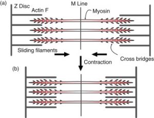
Sliding filament contraction model
In the sliding filament model, the thick and thin filaments cross, shortening the sarcomere. The movement often requires a skeletal muscle contraction, as can be seen when the biceps muscle of the upper arm contracts, pulling the forearm toward the trunk. The sliding filament model describes the process used by muscles to contract. It is a cycle of repetitive events that causes actin and myosin myofilaments to slide past each other, contracting the sarcomere and creating tension in the muscle.
Sarcomere structure
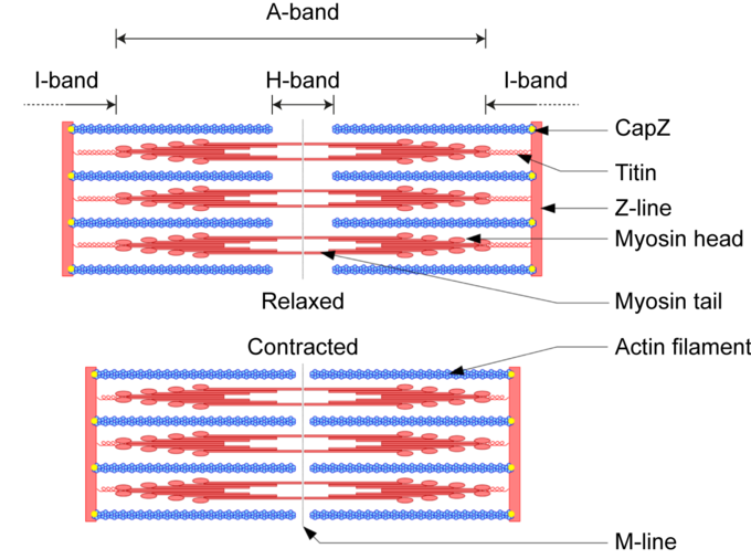
Understanding the sliding filament model requires an understanding of the structure of the sarcomere. A sarcomere is defined as the segment between two neighbouring parallel Z lines. Z lines are composed of a mixture of actin myofilaments and molecules of the highly elastic protein titin cross-linked by alpha-actinin. Actin myofilaments bind directly to the Z lines, while myosin myofilaments bind via titin molecules. Surrounding the Z line is the I band, the region where actin myofilaments do not overlap with myosin myofilaments. The I band is traversed by the titin molecule that connects the Z line to a myosin filament.
The region between two neighbouring parallel I bands is known as the A band and contains the full length of individual myosin myofilaments. Within the A band is a region known as the H band, which is the region that is not overlapped by actin myofilaments. Within the H band is the M line, which is composed of myosin myofilaments and titin molecules cross-linked by myomesin.
Titin molecules connect the Z line with the M line and provide a scaffolding for the myosin myofilaments. Its elasticity provides the basis for muscle contraction. Titin molecules are thought to play a key role as molecular ruler that maintains parallel alignment within the sarcomere. Another protein, nebulin, is thought to play a similar role in actin myofilaments.
Contraction Model
The molecular mechanism by which myosin and active myofilaments slide past each other is called the cross-bridge cycle. During muscle contraction, the myosin myofilament heads rapidly attach and release in a ratcheting fashion, pulling themselves along the actin myofilament. At the level of the sliding filament model, expansion and contraction only occur within the I and H bands. The myofilaments themselves do not contract or expand, so the A band remains constant.
The sarcomere and the sliding filament model of contraction: During contraction, myosin moves along actin myofilaments and compresses the I and H bands. During the stretch, this tension is released and the I and H bands expand. The A-band remains constant at all times since the length of the myosin myofilaments does not change. The amount of force and motion generated by an individual sarcomere is small. However, when multiplied by the number of sarcomeres in a myofibril, myofibrils in a myocyte, and myocytes in a muscle, the amount of force and motion generated is significant.